
Skip to content
Our promiseCare you can trust
Modern, Trusted DentistryHonest Advice. Your Best InterestConfidence in Your SmileClear Pricing. No Surprises

Age May be a Factor to Having Dental Implants. What THAT age is may surprise you!
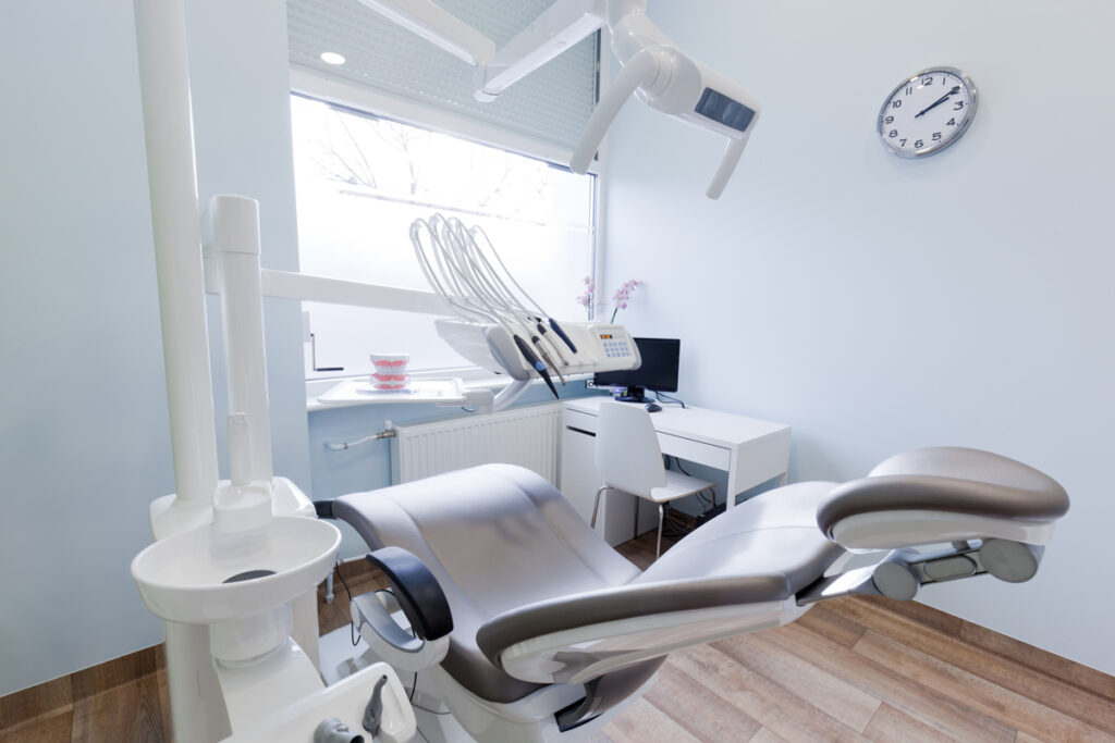
Anyone who wants to replace missing teeth are finding dental implants to be the best choice. Dental implants are the closest thing to natural teeth and the only procedure that doesn’t disrupt your existing teeth and facial structure.

By keeping your own structure, you can then eat, speak, laugh, and smile in confidence.

There are very few restrictions that may prevent a patient from receiving dental implants. In some instances, age may be one of the restrictions. Here’s what you need to consider:

Age restrictions regarding dental implants

  • Senior Citizens – You may think it would be the senior age group who might be the highly restricted group. But that would not be correct. In fact, dental implants have shown to be successful in patients in their 80s and even 90s. Though a patient’s health should be taken into consideration, there is generally no upper age limit for dental implant surgery. In fact, at least half of Americans over the age of 65 have at least one missing tooth. It would make sense people in this age group are good candidates for dental implants.
  • The Younger Patients – Odd as it may seem, it’s the younger age group that faces restrictions when it comes to dental implants. The procedure requires a fully developed jaw, so generally, a dental implant patient must be at least 17 or 18 years old. But this is not a set number. Every individual is different, and an oral surgeon can determine if a younger candidate is right for a dental implant procedure by taking x rays (often of the wrist) to see if the level of development is sufficient for surgery.

The fact is, a large number of people fall into the category of between the age of 18 and infinity. Should you want to consider getting implants, the best way to know if it’s right for you, is to talk to your dentist.

I’ll be happy to answer your questions or have you come in and give you a FREE Dental Implant & Healthy Smile Consultation without any obligation.

Call us at (281) 367-3685 in Woodlands , (281) 356-3351 in Pinehurst or CLICK HERE to schedule an appointment.

Accidental Displacement of Lower Third Molar – A Case Report

A 20 year old male patient reports-with the chief complaint of recurrent pain and swelling on the lingual side of the mandibular third molar area on the right side. The dental history of the patient revealed that lower right third molar was extracted by a local dentist and following this extraction, after about period of two weeks, the patient developed pain and swelling on the lingual side of the ramus. No further detailed dental history was available either from the patient or from the treating dentist. There were no clinical symptoms of dysesthesia of the lower lip or tongue on right side. The medical history of the patient was non-contributory.

CLINICAL EXAMINATION

The clinical examination was essentially restricted to intra-oral examination only. The intra-oral clinical examination revealed an absent lower third molar and tenderness on lingual side of lower third molar area on palpation.

RADIOLOGICAL EXAMINATION

A lateral oblique view of the mandible (Right Side) was taken and it showed an incompletely developed third molar in an unusual position raising doubts about its real position- in the ramus or in the surrounding soft tissues. A CT scan was ordered and the tooth was found lying lingual to the ramus below the mylohyoid muscle.

Considering the fact that the displaced tooth was causing pain and swelling because of infection, a decision was made to retrieve the displaced tooth. Reports of the routine preoperative blood and urine analysis were within normal limits.

SURGICAL PROCEDURE

The procedure was performed under local anaesthesia supplemented with premedication of Injectable Atropine (0.6 mg) and injectable Dexamethasomne (8 mg) given intra-muscularly and an oral tablet of Triazolam (0.5mg) given half an hour before surgery. A broad mucoperiosteal flap extending from the ramus to the distal surface of first pre-molar was raised on lingual side, the displaced tooth was pushed below the mylohyoid muscle which projected into the incision. Fibers of the mylohyoid muscle were split through blunt dissection, exposing the tooth. The tooth was grasped with a hemostat and delivered out of the mouth. The wound was irrigated with the copious amount of saline and antibiotic of Betadine (povidone-iodine 1%). Bleeding was arrested and subsequently, the wound was closed by interrupted black silk sutures. The patient was put on a course of an antibiotic (Cap. Amoxicillin 500mg.q8h) and an analgesic (Tab. Ibuproffen 400mg q8h) for a period of seven days. The postoperative course was uneventful. The surgical specimen of the retrieved displaced tooth was found to be non-carious tooth with incompletely formed roots.

Intra-Oral Sub-Mucosal Lipoma in the Mental Nerve Region – A Case Report And Review Of Literature

The World Health Organization classified lipoma and lipomatous lesions as follows in the year 2002 based on histopathological appearance of the lesions.

BENIGN LIPOMATOUS TUMORS

LIPOMA: A lipoma is a benign tumor composed of mature white adipocytes and is the most common soft tissue mesenchymal neoplasm in adults, usually a circumscribed, non-encapsulated aggregate with clear cytoplasm, absence of vascularity and atypia of metaplasia.

LIPOMATOSIS: (Madelung disease, Launois-Bensaude syndrome): Lipomatosis is a diffuse overgrowth of mature adipose tissue. It occurs in a variety of clinical settings and can affect different anatomic regions of the body. Diffuse lipomatosis usually occurs in individuals under 2 years of age but it may also arise in adults.

ANGIOLIPOMA: A subcutaneous nodule consisting of mature fat cells, intermingled with small and thin-walled vessels, a number if which contain fibrin thrombi. Angiolipomas most frequently present as multiple subcutaneous small nodules, usually tender or painful. There is no correlation between the intensity/occurrence of pain and the degree of vascularity.

LIPOBLASTOMA/ LIPOBLASTOMATOSIS: Lipoblastoma is a type of subcutaneous benign fatty tumor. Types include benign lipoblastomatosis, a tumor also knows as an embryonic lipoma, which usually occurs in children under three years old. This is a tumor of brown fat cells. The extremities are most commonly involved, but locations in the mediastinum, retroperitenoeum, trunk, head and neck, and various organs (lung, hear, parotic gland) have been described.

LIPOMATOSIS OF NERVES: Lipomatosis of nerve is characterized by infiltration of the epineurium by adipose and fibrous tissue. The tissue grows between and around nerve bundles thereby causing enlargement of the affected nerve. Patients present with a gradually enlarging mass in the affected area that may be asymptomatic or associated with motor 0r sensory deficits. Patients with macrodactyly have symmetrical or asymmetrical enlargement of the affected finger(s) with enlargement of the involved bones. Imaging studies show fusiform enlargement of the nerve with fatty infiltration.

HIBERNOMA: Hibernoma is a rare benign adipose tumor composed at least in part of brown fat cells with granular, multivacuolated cytoplasm. This brown gat component is admixed in variable proportion with white adipose tissue. Residual brown fat, mostly seen around cervical and axillary lymph nodes, should not be classified as hibernoma, Hibernoma is a relatively slow growing tumor of the subcutaneous tissue. At least 10% of cases are intramuscular. Hibernomas are usually painless. MRI reveals non-fat septations in hibernoma, not found in lipoma. On CT scan, hibernoma has a tissue attenuation intermediate between fat and skeletal muscle and is enhanced with contrast.

MYOLIPOMA: Myolipoma of soft tissue is a benign tumor exhibiting features of mature smooth muscle and mature adipose tissue. The majority of cases are deeply located and involve the abdominal cavity retroperitoneum, and inguinal areas. The trunk wall and extremities may also be involved; such cases are subcutaneous and may grow deeply to involve the superficial muscular fascia.

Treatment of Lipomas

NON EXCISIONAL TECHNIQUES

Non-excisional treatment of lipomas, which is now common, includes steroid injections and liposuction.

Steroid injections result in local fat atrophy, thus shrinking (or, rarely, eliminating) the lipoma. Injections are best performed on lipomas less than 1 inch in diameter. A one-to-one mixture of 1 percent lidocaine (Xylocaine) and triamcinolone acetonide (Kenalog), in a dosage of 10 mg per Ml, is injected into the center of the lesion. This procedure may be repeated several times at monthly intervals. The volume of steroid depends on the size of the lipoma, with an average of 1 to 3 mL of total volume administered. The number of injections depends on the response, which is expected to occur within three to four weeks. Complications, which are rare, are the result of the medication or the procedure, and can be prevented by injecting the smallest total amount possible and by positioning the needle so that it is in the center of the lipoma.

Liposuction can be used to remove small or large lipomatous growths, particularly those in locations where large scars should be avoided. Complete elimination of the growth is difficult to achieve with liposuction. Office procedure using a 16-gauge needle and a large syringe may be safer than large-cannula liposuction. Diluted lidocaine usually provides adequate anaesthesia for office liposuction.

ENUCLEATION:

Small lipomas can be removed by enucleation. A 3-mm to 4-mm incision is made over the lipoma. A curette is placed inside the wound and used to free the lipoma from the surrounding tissue, Once freed, the tumor is enucleated through the incision using the curette. Suctures generally are not needed, and a pressure dressing is applied to prevent hematoma formation.

EXCISION:

Larger lipomas are best removed through incisions made in the skin overlying the lipoma. The incisions are configured like a fusiform excision following the skin tension lines and are smaller than the underlying tumor. The central island of skin to be excised is grasped with a hemostat, or Allis clamp, which is used to provide traction for the removal of the tumor. Dissection is then performed beneath the subcutaneous fat to the tumor. Any tissue cutting is performed under direct visualization using a #15 scalpel or scissors around the lipoma. Care must be taken to avoid nerves or blood vessels that may lie just beneath the tumor.

The microscopic subtypes have no clinical significance with respect to the behaviour of tumors. Infiltrating lipomas are an exception, as the absence of a capsule and their ability to infiltrate the surrounding skeletal muscles represent a high-risk factor for recurrence. Depending upon the site, lipomas can be categorized into superficial, deep and periosteal. Although rare, the malignant transformation of oral lipomas into liposarcomas has been reported. Intramuscular location seems to be a risk factor for malignancy. Immunohistochemical detection of a aP2, a protein expressed by lipoblasts, could assist in differential diagnosis.

A histological examination after removal of lipomas is imperative to exclude lipo sarcomatous degeneration, to detect the absence of a capsule, which requires a constant follow-up due to a high probability of recurrence. Furthermore, the small number of reviews regarding lipomatous tumors of the oral cavity suggests the necessity of more attention on the soft tissue tumors that could affect the oral cavity. Thus, a possible expansion of the subtype classification and description if topographic issues may be revised in the future.


Find an office

LET’S MAKE YOU FEEL AT HOME

Plan your
next visit.

Choose your dental home to start your visit enquiry, or call the office directly.

Continue to visit form

Call (281) 356-3351

Submitting an enquiry does not confirm an appointment. The office will contact you.